Clinical case report · Kuala Lumpur
Optimizing wound bed preparation using a superoxidised solution and honey-based antimicrobial dressing in a Stage 3 sacral pressure injury: a clinical case report.
From wound-bed burden to healthy granulation.
Four illustrated checkpoints reveal the true post-debridement cavity and the healing response to revised care.
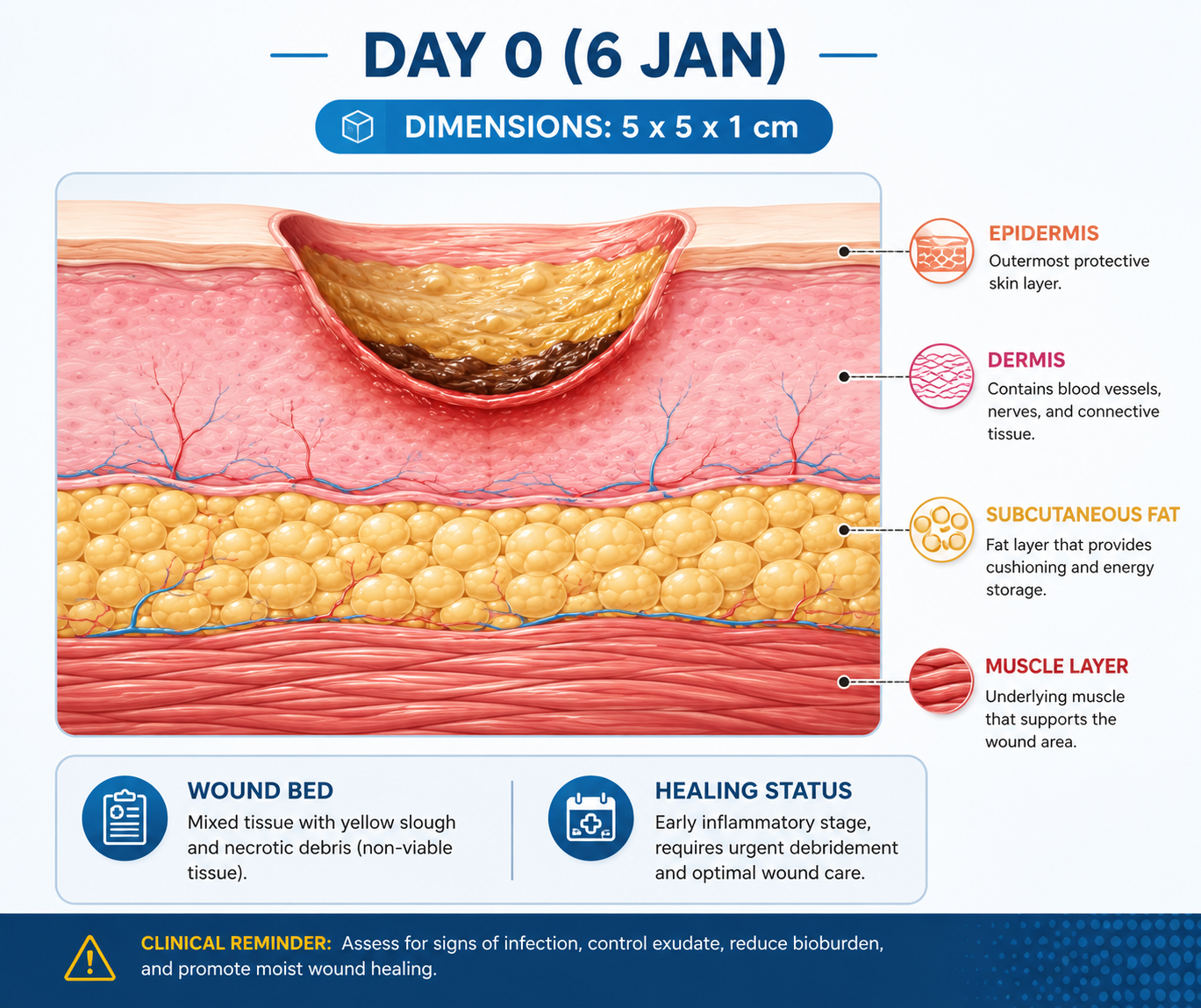
Slough and necrotic tissue · early granulation
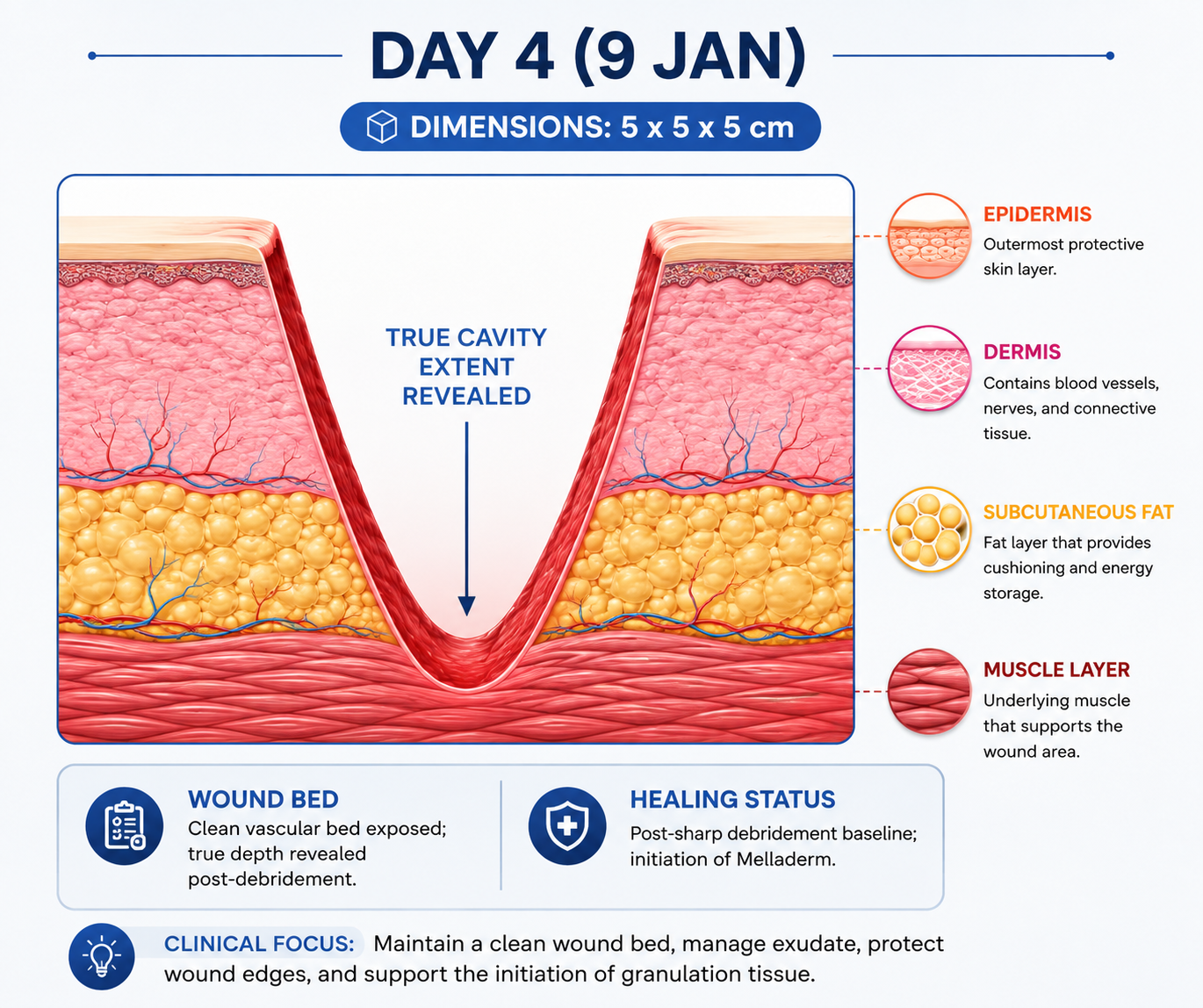
Post-sharp debridement · true cavity exposed
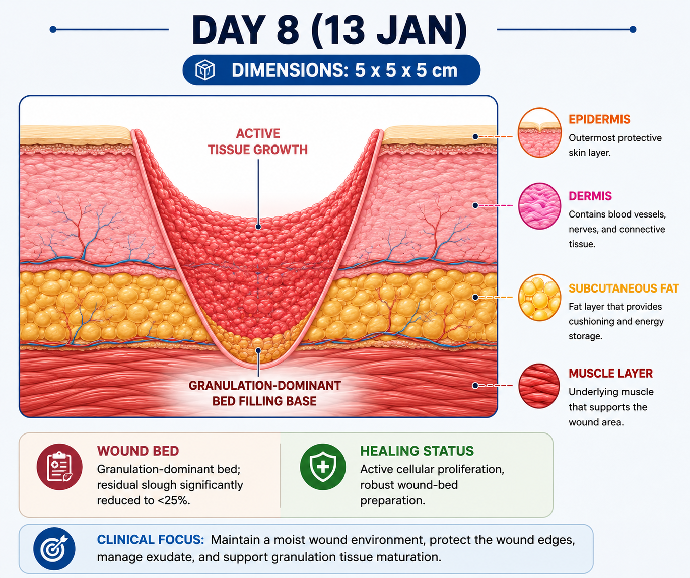
Granulation dominant · residual slough <25%
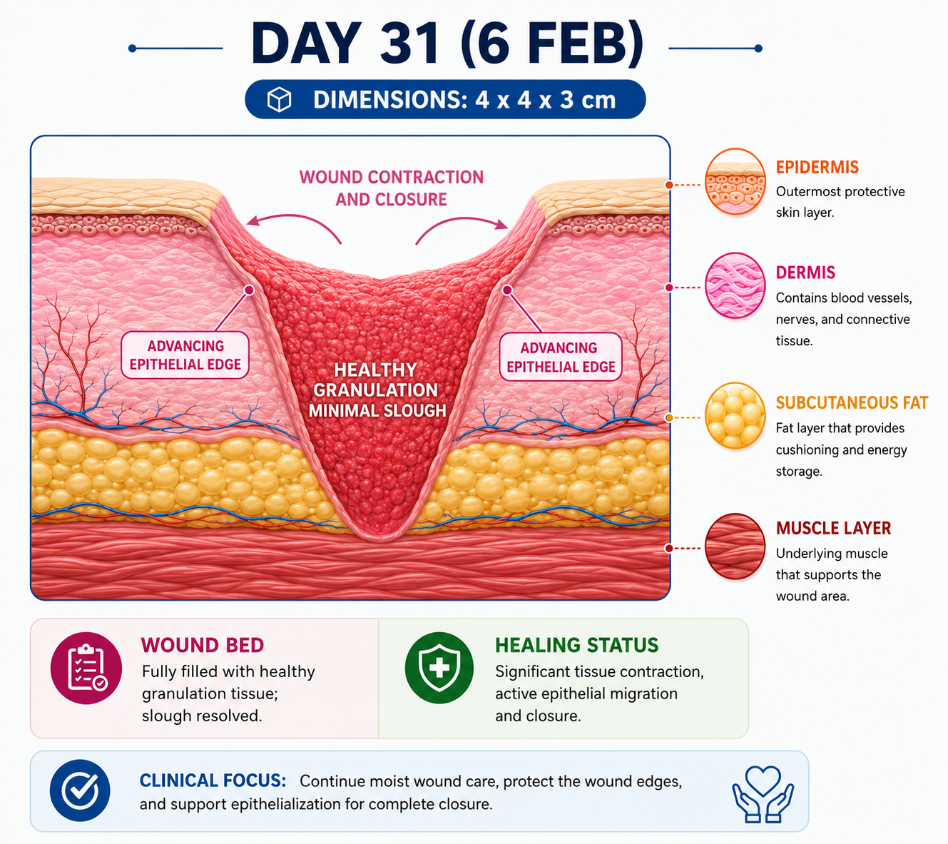
Healthy granulation · contraction and epithelialisation
The numbers show progressive healing.
Day 4 is the post-debridement baseline. Increased depth reflects exposure of the true cavity—not deterioration.
reduction between the Day 4 baseline and Day 31
Five interventions. One coordinated pathway.
Alternate-day care was adjusted to wound status and implemented by a multidisciplinary team.
True depth revealed. Healing achieved.
Post-debridement depth reflected exposure of the true wound cavity, not deterioration. TIME-based wound care and offloading achieved a 36% reduction in wound area and a 61.6% reduction in estimated cavity volume.
- Bus, S.A., et al. (2020) ‘Guidelines on offloading foot ulcers in persons with diabetes’, Diabetes/Metabolism Research and Reviews, 36(S1), e3274. doi:10.1002/dmrr.3274. (Offloading)
- Guo, S. and DiPietro, L.A. (2019) ‘Factors affecting wound healing’, Journal of Dental Research, 98(3), pp. 264–272. doi:10.1177/0022034518809361. (Anatomy and physiology of wound healing)
- Gottrup, F. (2020) ‘A new conceptual framework for wound dressings’, Advances in Wound Care, 9(2), pp. 77–84. doi:10.1089/wound.2019.1000.
- Schultz, G.S., et al. (2019) ‘Advancing the science of wound bed preparation: TIME’, International Wound Journal, 16(Suppl 1), pp. 1–9. doi:10.1111/iwj.13142. (Wound assessment & TIME concept)
- Sibbald, R.G., et al. (2021) ‘Wound bed preparation 2021’, Advances in Skin & Wound Care, 34(4), pp. 183–195. doi:10.1097/01.ASW.0000733725.68741.1c. (Wound cleansing and debridement)